Multiphoton microscopy (MPM) uses label-free contrast through optical signals generated by nonlinear near-infrared laser interactions with skin molecular components. Our goal is to use this unique molecular contrast to non-invasively diagnose skin diseases including skin cancer, to monitor the effects of skin treatments and to understand skin biology and functionality.
In vivo MPM of normal human skin [pub]
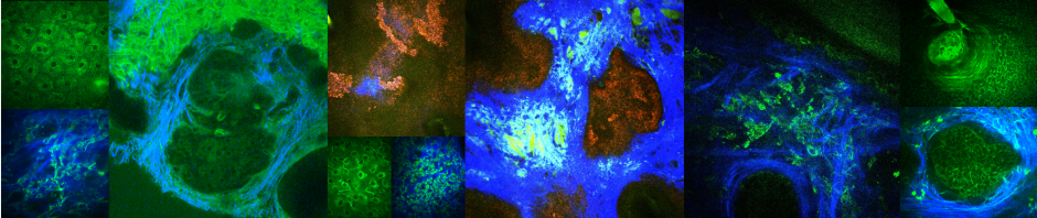
The contrast mechanisms of MPM in skin are based on the following signals:
Two-photon excited fluorescence from keratin, NADH/FAD, melanin, elastin fibers
Second harmonic generation from collagen fibers
Time-resolved single photon counting detection can provide additional sources of contrast.
Basal Cell Carcinoma (BCC)
BCC is a form of skin cancer originating from the basal cell layer of the epidermis and associated follicular structures. It is the most common human cancer, accounting for 25% of all cancer cases and 75% of skin malignant neoplasms diagnosed in the United States. BCC is diagnosed primarily by clinical evaluation and skin biopsy followed by sample preparation and histopathologic examination. We employ in vivo MPM imaging to evaluate the potential of this technology to be used for noninvasive histopathologic examination, a pain-free process that would be appreciated by patients and reduce the time from consultation to treatment.

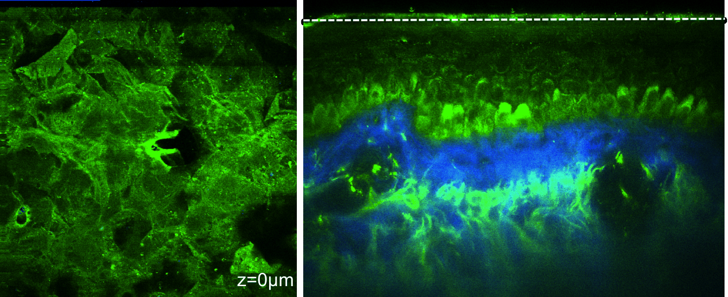
In vivo MPM can visualize nests of basaloid cells of BCC tumor in human skin
Clinical Collaborators: Christopher Zachary, MD; Kristen Kelly, MD; Ronald Harris, MD