
Radiotherapy is a critical component in the treatment of breast cancers (1). During radiation therapy, patients develop varying degrees of erythema with some developing dry or moist desquamation. Additionally, patients can develop permanent discoloration of the skin and thickening of the breast tissue (2). Quantifying radiation-induced skin changes during and after radiation is challenging, since standardized scoring systems (RTOG Radiation Morbidity Scoring Scheme and CTCAE v.4) are subjective and provide only a qualitative evaluation of the skins visual appearance (3). Additionally, there is currently no predictor for patients who develop more severe Grade 3 and 4 acute and late toxicities. Having a non-invasive quantitative technique to reproducibly and objectively measure acute and late radiation-induced skin changes is needed in the clinic.
The objectives of this study are to characterize radiation-induced skin changes during and after whole breast radiation using a contact-less operator-independent optical imaging device, Spatial Frequency Domain Imaging (SFDI). A reliable and quantitative method of risk assessment for radiation induced skin reactions would help in individualizing the management and care of patients undergoing radiotherapy. In the long term, it could also provide oncologists with a relatively simple, risk-free bedside tool that can help inform medical decisions on radiation protocols (such as dose, frequency and duration) thereby minimizing unnecessary skin toxicity while providing adequate treatment efficacy.
Presented at Military Health System Research Symposium, August 2018
Authors
Anaïs Leproux1, Randy Wei2, Jeffrey Kuo3, Parima Daroui3, Nilam Ramsinghani3, Muthana Al-Ghazi3 and Anthony J Durkin1,4*
1Beckman Laser Institute and Medical Clinic, University of California Irvine, CA 92612, USA
2Memorial Radiation Oncology Medical Group, 18111 Brookhurst Street, Suite LL0300, Fountain Valley, CA 92708
3Department of Radiation Oncology, 101 The City Drive S, Orange, CA 92868
4Department of Biomedical Engineering, University of California, Irvine, California 92697, USA
Methods
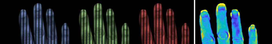
SFDI is a non-invasive and rapid functional imaging technique. It uses spatially-structured light at 8 wavelengths (from 470 to 850 nm) to quantitatively map superficial tissue absorption and scattering in a wide-field, non-contact imaging geometry. Tissue oxygen saturation, oxyhemoglobin, deoxyhemoglobin, and melanin content can be calculated from the SFDI absorption data.
Fourteen female breast cancer patients prescribed for radiation therapy were enrolled in in this study under a clinical protocol approved by the Institutional Review Board at the University of California, Irvine. SFDI measurements were performed before the start of treatment, weekly during radiation treatment and 2 weeks, 1.5 months, 3 months and 6 months after completion of treatment. The contralateral, untreated breast was also measured and used as an internal control.
Results
Increased melanin content was observed over the treated area, correlating with hyperpigmentation. Subsequent decrease in melanin content was observed in patients who experienced skin discoloration. Increase in total hemoglobin was consistently observed over the treated area, correlating with erythema. A drop in tissue oxygen saturation was observed in some patients at about 4 or 5 weeks of treatment, probably resulting from radiation-induced damage of the microvasculature. This drop was preceded by a steady increase in oxygen saturation. Normalization of the total hemoglobin, saturation and melanin were observed at the post-radiation time-points, suggesting healing of the skin.
Conclusions
Our preliminary data in fourteen (14) subjects have validated the feasibility and reproducibility of SFDI to measure spatial and temporal changes in the skin during and after radiation therapy. Potentially, SFDI could be used to measure different prophylactic creams during radiation treatment, or predict severity of radiation skin changes from pre-treatment SFDI measurements.
Acknowledgements
We thankfully recognize support from the NIH, including NIGMS grant R01GM108634 and NIBIB P41EB015890 (A Biomedical Technology Resource). We also thank the Arnold Beckman Foundation. Any opinions, finding, and conclusions or recommendations expressed in this material are those of the authors and do not necessarily reflect the views of the organizations above.
References
- Margolese RG, Hortobagyi GN, Buchholz TA. The Role of Radiation for invasive Breast Cancer2003.
- White J, Joiner MC. Toxicity from Radiation in Breast Cancer. In: Small W, Woloschak GE, editors. Radiation Toxicity: A Practical Guide. Boston, MA: Springer US; 2006. p. 65-109.
- López E, Núñez MI, Guerrero MR, del Moral R, de Dios Luna J, del Mar Rodríguez M, Valenzuela MT, Villalobos M, de Almodóvar JMR. Breast cancer acute radiotherapy morbidity evaluated by different scoring systems. Breast Cancer Research and Treatment. 2002;73(2):127-34.