
Cardiac arrest (CA) affects over 565,000 people annually in the United States [1]. Survivors usually experience some form of neurological damage due to loss of oxygen supply to the brain. In the intensive care unit, cerebral oxygen metabolism is measured, by removing samples of blood from the jugular vein. This technique does not provide real-time feedback to guide clinicians in improving neurological outcomes during and following cardiac arrest.

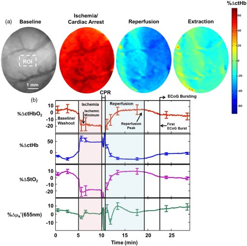
In order to understand the brain’s response to cardiac arrest, optical imaging methods could be used. Optical techniques such as Laser Speckle Imaging (LSI) and Spatial Frequency Domain Imaging (SFDI) provide quantitative information about the cerebral blood flow (CBF) and oxygenation of the brain. These techniques are helpful for the following:
- No well validated clinical treatments are available for improving patient outcome following CA.
- 9% of survivors walk out the hospital with good neurological performance [3].
- Cardiopulmonary Resuscitation (CPR) can often only supply oxygen to certain parts of the brain [2].
- Optical imaging can acquire information over the entire region of the brain that is exposed to the camera.
The WiFI team collaborated with Dr. Yama Akbari and his lab. Dr. Akbari is a physician scientist who focuses on neuro-intensive care using basic science, translational, and clinical approaches. His lab specializes in brain monitoring electrocorticography (ECoG) signals. The combination of optical imaging and brain monitoring will help better understand the response to different clinical interventions following CA and CPR.
Authors


Collaborator:
Dr. Yama Akbari Lab, UC Irvine Department of NeurologyCitation
High-speed spatial frequency domain imaging of rat cortex detects dynamic optical and physiological properties following cardiac arrest and resuscitation.
Wilson, R. H., Crouzet, C., Torabzadeh, M., Bazrafkan, A. K., Hosseini-Farahabadi, M., Jamasian, B., … Tromberg, B. J. (2017). Neurophotonics, 4(4), 45008–45009.Cerebral blood flow is decoupled from blood pressure and linked to EEG bursting after resuscitation from cardiac arrest.
Crouzet, C., Wilson, R. H., Bazrafkan, A., Farahabadi, M. H., Lee, D., Alcocer, J., … Akbari, Y. (2016). Biomedical Optics Express, 7(11), 4660–4673.