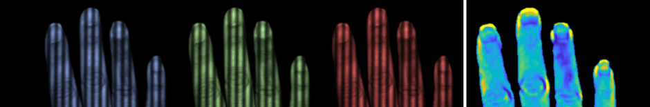
We have developed compressed sensing single pixel spatial frequency domain imaging (cs-SFDI) to characterize tissue optical properties over a wide field of view ( 35 mm × 35 mm ) using multiple near-infrared (NIR) wavelengths simultaneously. Our approach takes advantage of the relatively sparse spatial content required for mapping tissue optical properties at length scales comparable to the transport scattering length in tissue ( μ tr ∼ 1 mm ) and the high bandwidth available for spectral encoding using a single-element detector. cs-SFDI recovered absorption ( μ a ) and reduced scattering ( μ s ′ ) coefficients of a tissue phantom at three NIR wavelengths (660, 850, and 940 nm) within 7.6% and 4.3% of absolute values determined using camera-based SFDI, respectively. These results suggest that cs-SFDI can be developed as a multi- and hyperspectral imaging modality for quantitative, dynamic imaging of tissue optical and physiological properties.Citation
Compressed single pixel imaging in the spatial frequency kingdom.
Mohammad Torabzadeh, Il-Yong Park, Randy A. Bartels, Anthony J. Durkin, Bruce J. Tromberg. J. Biomed. Opt. 22(3) 030501 (16 March 2017)