Hair loss is frequently underestimated in general medical practice, although it causes a significant impact in the quality of life of patients. Traditionally, hair loss has been classified into scarring and non-scarring conditions. In non-scarring alopecia, hair follicles are preserved with potential for hair regrowth. In scarring alopecia, the hair follicle is irreversibly destroyed due to destruction of stem cells in the bulge area of the outer root sheath, and replaced by fibrous scar tissue, leading to permanent hair loss. Accurate diagnosis is important for optimal treatment and management of alopecia and can be challenging due to the limitations of histologic studies and the side effects of scalp biopsies. In a recent study, we evaluated in the ability of the MPM imaging technology to non-invasively identify morphological features that can distinguish scarring from non-scarring alopecia in human skin.

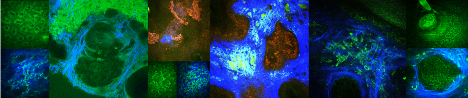
MPM can visualize inflammation in scarring alopecia of human skin. (A) Clinical image of temporal scalp area affected by scarring alopecia. (B) Dermoscopic image of frontal scalp area in A. (C–F) MPM images acquired at different depths showing an almost obliterated empty hair follicle with no hair shaft. Sebaceous glands are notably absent. Keratinocytes are visible in the stratum granulosum and stratum spinosum layers (z=40 um and z=50 um). Inflammatory cells (lymphocytes and macrophages, orange arrows in C–F), elastin and collagen fibers surround the follicle. Scale bar is 20 um. (G)A 3D view of MPM images in a z-stack from which the (C–F) images were selected.
Clinical collaborator: Natasha Mesinkovska, MD, PhD