Skin Cancer
Melanoma
Although the overall cancer incidence rates decreased among men and remained stable among women over the past 5 years of available data (2012-2016), the melanoma of the skin incidence shows a high increase rate compared to other cancers for both men and women. Early detection remains difficult but is critical for successful treatment. We employ in vivo MPM imaging to evaluate the potential of this technology to non-invasively characterize and diagnose pigmented lesions suspected of melanoma.

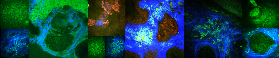
MPM can visualize key melanoma signature features: migration of melanocytes in upper epidermal layers (arrows), proliferation of atypical melanocytes at the dermo-epidermal junction and presence of melanoma cells and melanophages in the papillary dermis.
Clinical Collaborators: Kristen Kelly, MD; Christopher Zachary, MD; Ronald Harris, MD; Ronald Barr, MD; Janellen Smith, MD; Ken Linden, MD; Anand Ganesan, MD, PhD
Related publications
Basal Cell Carcinoma (BCC)
BCC is a form of skin cancer originating from the basal cell layer of the epidermis and associated follicular structures. It is the most common human cancer, accounting for 25% of all cancer cases and 75% of skin malignant neoplasms diagnosed in the United States. BCC is diagnosed primarily by clinical evaluation and skin biopsy followed by sample preparation and histopathologic examination. We employ in vivo MPM imaging to evaluate the potential of this technology to be used for noninvasive histopathologic examination, a pain-free process that would be appreciated by patients and reduce the time from consultation to treatment.

In vivo MPM can visualize nests of basaloid cells of BCC tumor in human skin
Clinical Collaborators: Christopher Zachary, MD; Kristen Kelly, MD; Ronald Harris, MD