Melasma
Melasma is a skin disorder characterized by irregular light to dark brown macules frequently found on the sun-exposed areas of the face, due to increased melanin production and deposition. Identifying the depth of excess pigment (epidermal and/or dermal) is critical for successful treatment. To date, there is no reliable method to determine the depth of melanin pigment, a key factor among others that make the treatment of melasma a difficult challenge. Biopsy is not commonly an option due to cosmetic reasons, and a heterogeneous melanin distribution is often present, which would require multiple sampling sites. We employ in vivo MPM imaging to evaluate the potential of this technology to non-invasively characterize the melanin content, location, and distribution in melasma of human skin.

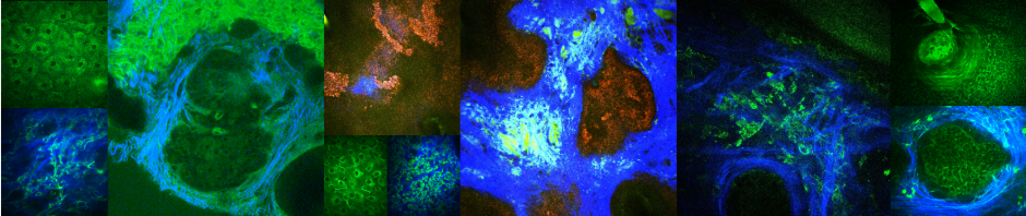
MPM can visualize dermal melanophages in melasma lesions of human skin- In vivo MPM images showing: (A) clusters of small dermal melanophages (arrows), (B) clusters of large dermal melanophages (arrows), (C) dendritic melanophages (arrows), and (D) individual/scattered dermal melanophages (arrow). Images in A, B, C are from subjects with dermal/mixed melasma. Image D corresponds to epidermal melasma. Collagen and elastin fibers are visualized in dermis by their SHG (blue) and TPEF (green) signals, respectively. ‘z’ represents depth in all MPM images. Scale bar is 40 μm.
Clinical collaborator: Anand K. Ganesan, MD